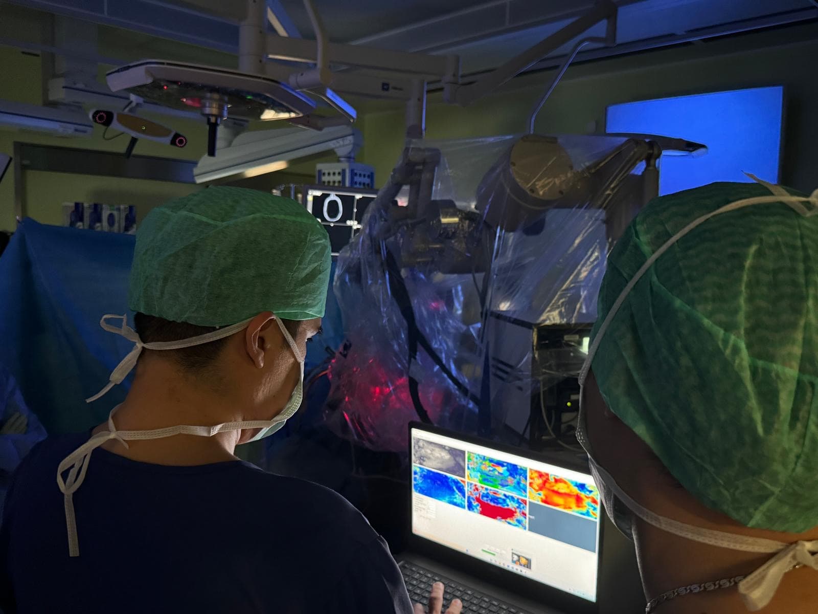
La chirurgie est l'étape décisive du traitement des cancers du cerveau ; pourtant, même avec les microscopes chirurgicaux modernes, la frontière entre tissu sain et tissu tumoral reste très difficile à voir. Le contraste visuel entre tumeur et tissu non tumoral est faible, mais il existe entre eux une nette différence structurelle. La matière blanche du cerveau sain, composée de faisceaux densément empilés d'axones myélinisés, constitue un milieu optiquement anisotrope. Les cellules tumorales croissent de façon chaotique et détruisent ou déplacent les faisceaux de fibres : l'anisotropie optique disparaît dans la zone tumorale. AOPLab conçoit et construit des systèmes de polarimétrie de Mueller à imagerie grand champ capables de visualiser l'anisotropie optique des tissus biologiques et de rendre visible en temps réel la frontière entre zones à fibres préservées et zones sans fibres. Cette modalité optique est sans marquage et ne perturbe pas le déroulement de l'intervention. Le prototype développé dans le cadre de notre précédent projet HORAO a été testé avec succès ex vivo et in vivo. Nous développons désormais la prochaine génération d'instruments polarimétriques, plus précis, plus compacts et plus rapides, pour la chirurgie du cerveau, du rachis, de la tête et du cou.
Le Défi Clinique
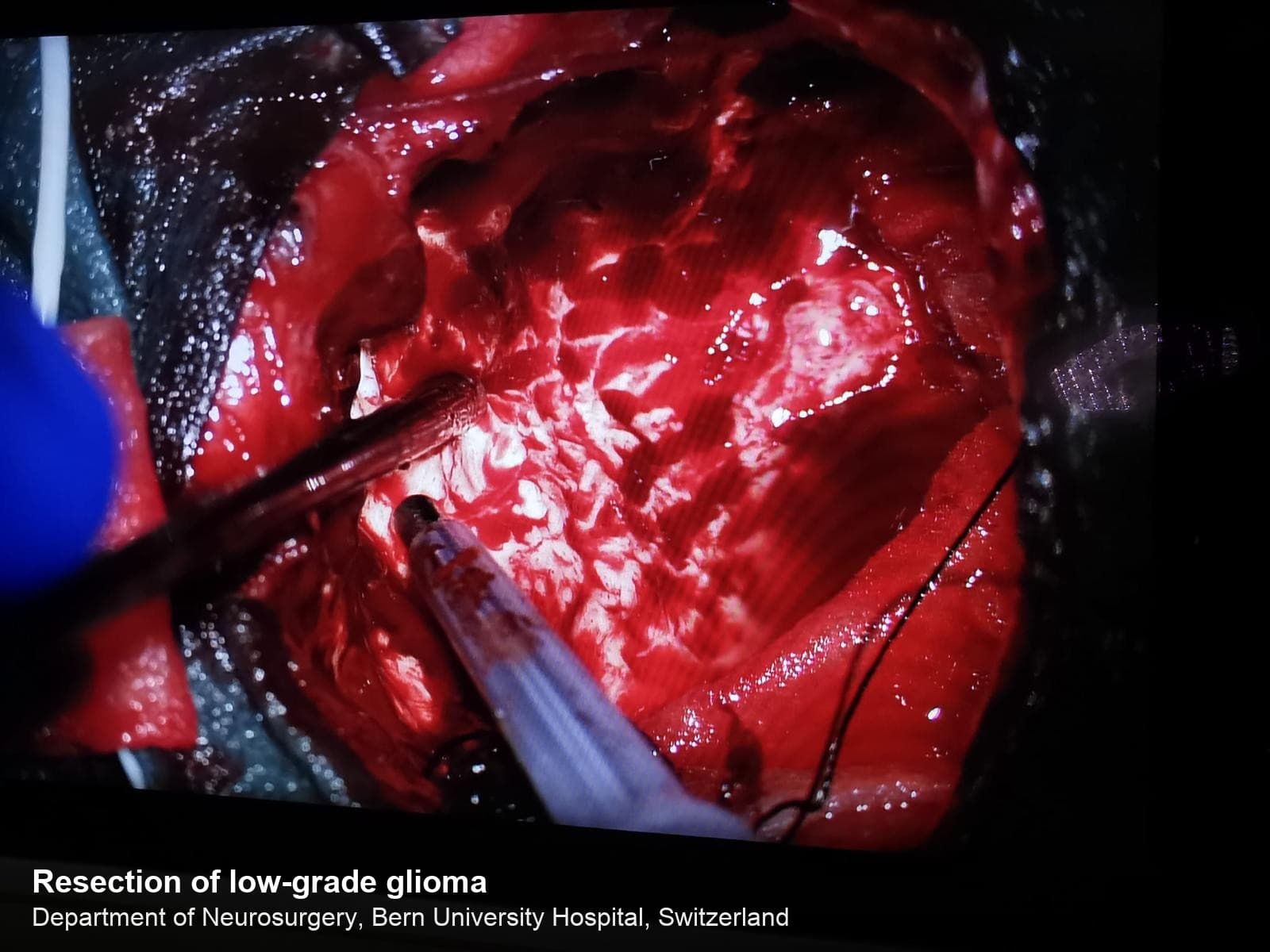
La chirurgie des tumeurs cérébrales recherche un équilibre délicat : retirer le plus de tissu tumoral possible tout en protégeant les faisceaux de fibres de la matière blanche, qui contrôlent le mouvement, la parole et la vision. Sous éclairage en lumière blanche, la frontière entre tumeur et tissu non tumoral est à peine visible : le chirurgien risque soit de laisser des cellules tumorales résiduelles (principale cause de récidive), soit d'endommager des faisceaux fonctionnels (principale cause de déficit neurologique postopératoire).
Voir l'Architecture Cachée de la Matière Blanche
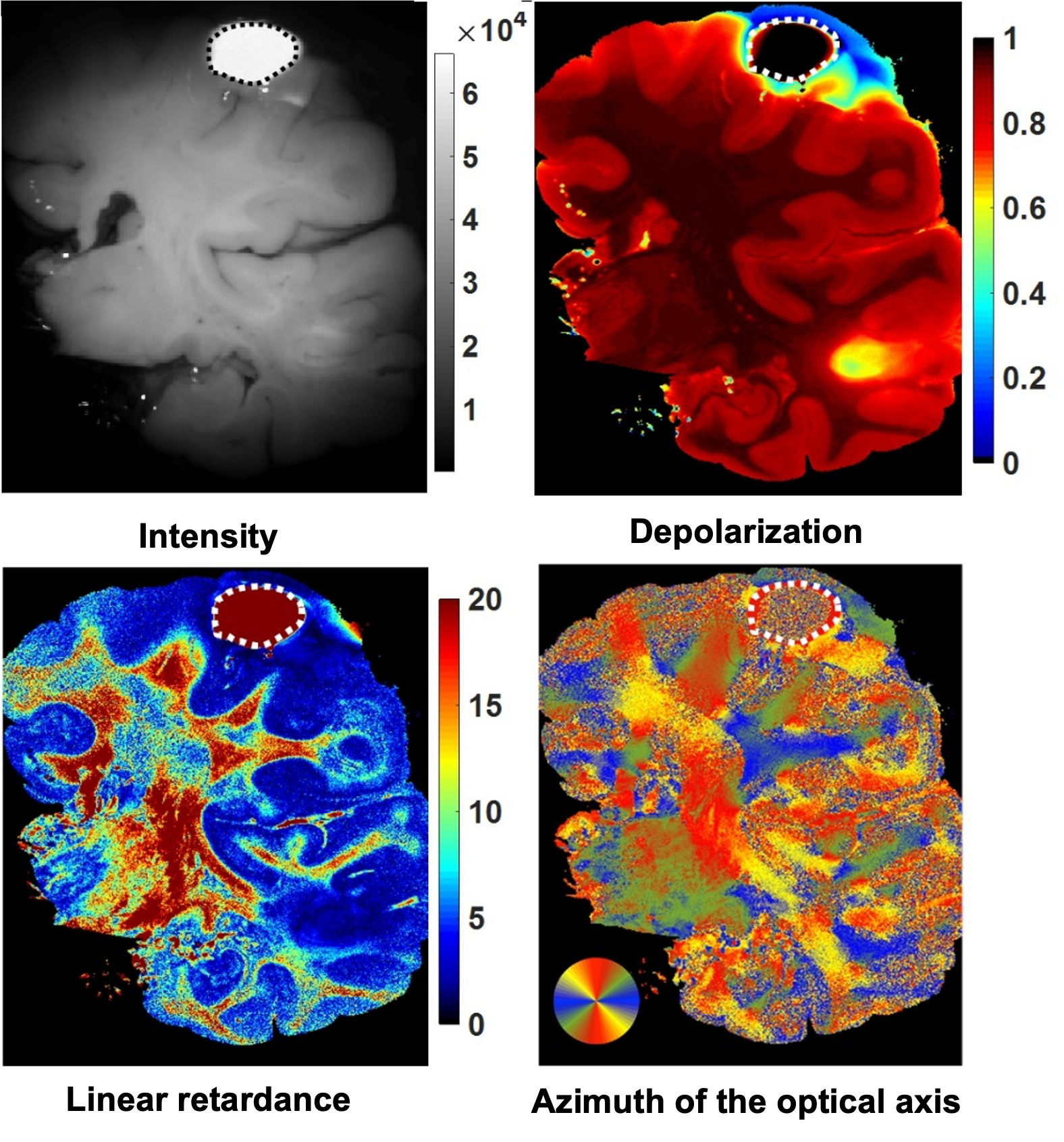
La matière blanche du cerveau sain est constituée de faisceaux alignés et densément empilés d'axones myélinisés, à l'origine d'une biréfringence dite de forme. L'axe optique de ce milieu linéairement biréfringent est corrélé à la direction des faisceaux de fibres. Notre polarimètre de Mueller à imagerie grand champ, conçu sur mesure, mesure les images complètes de la matrice de Mueller 4×4 et, par un algorithme non linéaire de compression des données appliqué pixel par pixel — la décomposition de Lu–Chipman (S.-Y. Lu et R. A. Chipman, J. Opt. Soc. Am. A 13(5), 1106–1113 (1996), 10.1364/JOSAA.13.001106) —, produit les cartes de dépolarisation, de retard linéaire et d'azimut de l'axe optique du tissu cérébral.
Distinguer la Tumeur Cérébrale du Tissu Non Tumoral
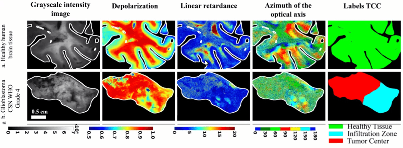
La croissance tumorale détruit l'architecture ordonnée de la matière blanche saine, et le tissu tumoral perd son anisotropie optique. Toutes les propriétés polarimétriques du cerveau sont affectées : les valeurs de dépolarisation et de retard linéaire chutent, et l'azimut de l'axe optique prend des valeurs aléatoires dans la zone tumorale. Sur des échantillons de tissu sain et de glioblastome (grade 4 OMS), les valeurs de dépolarisation et de retard suivent la transition du parenchyme sain, à travers la zone d'infiltration, jusqu'au cœur de la tumeur, en accord avec la vérité histologique. C'est le contraste structurel qui manque aux images en lumière blanche, et le signal qu'apprennent à exploiter nos modèles de segmentation.
Premières Mesures Polarimétriques du Cerveau In Vivo
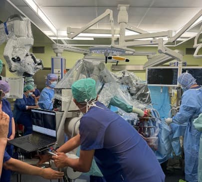
Après la validation de la chaîne d'imagerie polarimétrique sur des centaines d'échantillons ex vivo, un nouveau système polarimétrique de Mueller miniaturisé a été déployé lors de résections de tumeurs cérébrales, produisant les premières mesures polarimétriques in vivo de ce type. Cela marque le début du passage de la polarimétrie de Mueller à imagerie grand champ du banc optique au bloc opératoire, et une étape clé vers la visualisation sans marquage et en temps réel de la tumeur cérébrale et des faisceaux de fibres du tissu non tumoral pendant la neurochirurgie.
Related Publications
Gros, É., Sakiri, A., Moriconi, S., Jankauskas, A., Hasler, D., Lucas, T., McKinley, R., Maragkou, T., Novikova, T., Schucht, P., Rodríguez-Núñez, O.
Brain Tissue Polarimetry Under Simulated Intraoperative Conditions: Effects of Edema and Temperature
Journal of Innovative Optical Health Sciences·2026
Chae, S., Wang, M., Lucas, T., Huang, T., Albano, G., Rodríguez-Núñez, O., Pierangelo, A., Gros, É., Maragkou, T., McKinley, R., Schucht, P., Novikova, T.
Biomedical Optics Express·2026
Tuchin, V. V., Novikova, T., Wang, L. V., Zimnyakov, D. A., Ma, H., Alonova, M. V., Wan, J.
Optical Polarization in Biomedical Applications (2nd Edition)
Springer Series in Biophysics, Springer Berlin Heidelberg New York·2026
Novikova, T., Chae, S., Lucas, T., Rodríguez-Núñez, O.
Polarized-light microscopy
Optical Techniques in Clinical Applications, Springer Nature·2026
Wang, M., Chae, S., Lucas, T., Rodríguez-Núñez, O., Gros, É., Hahne, C., Maragkou, T., McKinley, R., Schucht, P., Novikova, T.
Proc. SPIE 13854, 1385405·2026
Diamant, A., Lucas, T., Rodríguez-Núñez, O., Chae, S., Gros, É., Hahne, C., Maragkou, T., McKinley, R., Schucht, P., Novikova, T.
Proc. SPIE 13854, 138540F·2026
Oberti, N., Rodríguez-Núñez, O., Hasler, D., Schucht, P., McKinley, R., Novikova, T., Maragkou, T., Gros, É.
Proc. SPIE 14098, Tissue Optics and Photonics IV, 140981H (SPIE Photonics Europe)·2026
Lucas, T., Rodríguez-Núñez, O., Iqbal, A., Chae, S., Gros, É., Hahne, C., Maragkou, T., McKinley, R., Schucht, P., Novikova, T.
Proc. SPIE PC14094, Biomedical Spectroscopy, Microscopy, and Imaging IV, PC1409405 (SPIE Photonics Europe)·2026
Rodríguez-Núñez, O., Gros, É., Lucas, T., Chae, S., Hahne, C., Hasler, D., Maragkou, T., McKinley, R., Novikova, T., Schucht, P.
Proc. SPIE PC13841, Multimodal Biomedical Imaging XXI, PC1384107 (SPIE Photonics West)·2026
Rodríguez-Núñez, O., Lucas, T., Gros, É., Baier, D., Chae, S., Hahne, C., Hasler, D., Maragkou, T., McKinley, R., Schucht, P., Novikova, T.
Proc. SPIE 13854, Polarized Light and Optical Angular Momentum for Biomedical Diagnostics 2026 (SPIE Photonics West)·2026
Wang, M., Chae, S., Lukinsone, V., Lucas, T., Rodríguez-Núñez, O., Gros, É., Hahne, C., Maragkou, T., McKinley, R., Schucht, P., Novikova, T.
Biomedical Optics Express, 16(12), 5261–5278·2025
Hahne, C., Rodríguez-Núñez, O., Gros, É., Lucas, T., Novikova, T., Maragkou, T., Schucht, P., McKinley, R.
Physically Consistent Image Augmentation for Deep Learning in Mueller Matrix Polarimetry
IEEE Transactions on Image Processing, 34, 6953–6962·2025
Hahne, C., Diaz, I., Rodríguez-Núñez, O., Gros, É., Lucas, T., Novikova, T., Maragkou, T., Schucht, P., McKinley, R.
Polarimetric Feature Analysis of Mueller Matrices for Brain Tumor Image Segmentation
Optics Express, 33(20), 43379·2025
Gros, É., Rodríguez-Núñez, O., Moriconi, S., McKinley, R., Hewer, E., Lucas, T., Vassella, E., Schucht, P., Novikova, T., Hahne, C., Maragkou, T.
Alignment of Histological and Polarimetric Large-Scale Imaging for Brain Tissue Characterization
Journal of Biomedical Optics, 30(9), 096003·2025
Novikova, T.
Imaging Mueller polarimetry for biomedical diagnosis: Recent developments
EPJ Web of Conferences, 335, 04007·2025
Novikova, T.
Imaging Mueller Polarimetry: a New Lens on Human Health
Optica Biophotonics Congress 2025, Technical Digest Series, paper TTu3F.1·2025
Maragkou, T., Gros, R., Rodríguez-Núñez, O., Felger, L., Moriconi, S., McKinley, R., Novikova, T., Schucht, P., Pierangelo, A., Vassella, E., Hewer, E.
Diagnosis of Brain Tumors Using Wide-Field Imaging Mueller Polarimetry
Journal of Neuropathology & Experimental Neurology, 83(6), 409·2024
Gros, R., Rodríguez-Núñez, O., Felger, L., Moriconi, S., McKinley, R., Pierangelo, A., Novikova, T., Vassella, E., Schucht, P., Hewer, E., Maragkou, T.
IEEE Transactions on Medical Imaging, 43(12), 4120·2024
Moriconi, S., Rodríguez-Núñez, O., Gros, R., Felger, L. A., Maragkou, T., Hewer, E., Pierangelo, A., Novikova, T., Schucht, P., McKinley, R.
Near-real-time Mueller polarimetric image processing for neurosurgical intervention
International Journal of Computer Assisted Radiology and Surgery·2024
Gros, R., Rodríguez-Núñez, O., Felger, L., Moriconi, S., McKinley, R., Pierangelo, A., Novikova, T., Vassella, E., Schucht, P., Hewer, E., Maragkou, T.
Proc. SPIE 13010, Tissue Optics and Photonics III, 130100F·2024
Ivanov, D., Si, L., Felger, L., Maragkou, T., Schucht, P., Ma, H., Schanne-Klein, M.-C., Ossikovski, R., Novikova, T.
Journal of Biomedical Optics, 28(10), 102908·2023
Gros, R., Rodríguez-Núñez, O., Felger, L., Moriconi, S., McKinley, R., Pierangelo, A., Novikova, T., Vassella, E., Schucht, P., Hewer, E., Maragkou, T.
Effects of formalin fixation on polarimetric properties of brain tissue: fresh or fixed?
Neurophotonics, 10(2), 025009·2023
Felger, L., Rodríguez-Núñez, O., Gros, R., Maragkou, T., McKinley, R., Moriconi, S., Murek, M., Zubak, I., Novikova, T., Pierangelo, A., Schucht, P.
Biomedical Optics Express, 14(5), 2400–2415·2023
Yashin, K., Novikova, T., Shcheslavskiy, V.
Editorial: Optical imaging and Laser technologies in Neuro-Oncology
Frontiers in Oncology, 12, 1103711·2023
Moriconi, S., Felger, L., Rodríguez-Núñez, O., Gros, R., Hewer, E., Maragkou, T., Novikova, T., Pierangelo, A., Murek, M., Schucht, P., McKinley, R.
Towards Real-time Integration of Polarimetric Image-processing for Neurosurgical Applications
Brain and Spine, 3, 102159·2023
Rodríguez-Núñez, O., Gros, R., Felger, L., Maragkou, T., McKinley, R., Moriconi, S., Novikova, T., Pierangelo, A., Schucht, P.
Polarimetric markers for delineation of lesional margins during brain tumor surgery
Brain and Spine, 3, 102160·2023
Felger, L., Gros, R., Maragkou, T., McKinley, R., Moriconi, S., Murek, M., Novikova, T., Pierangelo, A., Rodríguez-Núñez, O., Zubak, I., Schucht, P.
Proc. SPIE 12364, 1236406·2023
Gros, R., Felger, L., Maragkou, T., McKinley, R., Moriconi, S., Murek, M., Novikova, T., Rodríguez-Núñez, O., Schucht, P., Zubak, I., Pierangelo, A.
Proc. SPIE PC12382, PC1238203·2023
McKinley, R., Felger, L., Gros, R., Hewer, E., Maragkou, T., Moriconi, S., Murek, M., Novikova, T., Rodríguez-Núñez, O., Pierangelo, A., Schucht, P.
Proc. SPIE PC12382, PC1238208·2023
Moriconi, S., Felger, L., Gros, R., Hewer, E., Maragkou, T., Murek, M., Novikova, T., Rodríguez-Núñez, O., Pierangelo, A., Schucht, P., McKinley, R.
Denoising diffusion networks applied to Mueller polarimetric images of brain tissue
Proc. SPIE PC12382, PC123820H·2023
Rodríguez-Núñez, O., Felger, L., Gros, R., Maragkou, T., McKinley, R., Moriconi, S., Murek, M., Pierangelo, A., Schucht, P., Zubak, I., Novikova, T.
Proc. SPIE PC12382, PC1238209·2023
Novikova, T., Rodríguez-Núñez, O.
Polarimetric techniques for the structural studies and diagnosis of brain
Advanced Optical Technologies, 11(5-6), 157–171·2022
Novikova, T., Pierangelo, A., Schucht, P., Meglinski, I., Rodríguez-Núñez, O., Lee, H. R.
Mueller Polarimetry of Brain Tissues
Polarized Light in Biomedical Imaging and Sensing, Springer·2022
McKinley, R., Felger, L., Hewer, E., Maragkou, T., Murek, M., Novikova, T., Rodríguez-Núñez, O., Pierangelo, A., Schucht, P.
Proc. SPIE 12136, 121360G·2022
Rodríguez-Núñez, O., Schucht, P., Hewer, E., Novikova, T., Pierangelo, A.
Brain fiber tracts imaging with polarized light: a potential navigation tool for tumor neurosurgery
Proc. SPIE PC11963, PC1196303·2022
Rodríguez-Núñez, O., Schucht, P., Hewer, E., Novikova, T., Pierangelo, A.
Polarimetric visualization of healthy brain fiber tracts under adverse conditions: ex vivo studies
Biomedical Optics Express, 12(10), 6674–6685·2021
Novikova, T., Schucht, P., Lee, H. R., Mezouar, M. H., Hewer, E., Raabe, A., Murek, M., Zubak, I., Goldberg, J., Kövari, E., Pierangelo, A.
Proc. SPIE 11641, Dynamics and Fluctuations in Biomedical Photonics XVIII, 1164105·2021
Rodríguez-Núñez, O., Schucht, P., Lee, H. R., Mezouar, M. H., Hewer, E., Raabe, A., Murek, M., Zubak, I., Goldberg, J., Kövari, E., Pierangelo, A., Novikova, T.
Proc. SPIE 11919, Translational Biophotonics: Diagnostics and Therapeutics, 119190Y·2021
Schucht, P., Lee, H. R., Mezouar, M. H., Hewer, E., Raabe, A., Murek, M., Zubak, I., Goldberg, J., Kövari, E., Pierangelo, A., Novikova, T.
IEEE Transactions on Medical Imaging, 39(12), 4376–4382·2020