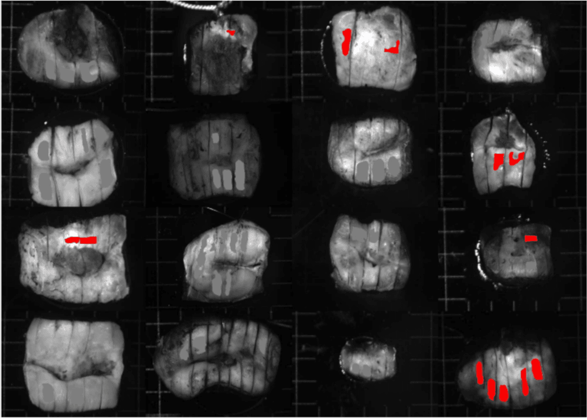
AOPLab explores the potential of wide-field imaging Mueller polarimetry to address two important gynecological problems, where the assessment of uterine cervix tissue microstructure holds key information.
The first problem is related to the early detection of cervical pre-cancer, where screening currently still relies on the results of Pap smear and on subjective colposcopic examination of the patient. Wide-field Mueller polarimetric imaging aims to classify cervical intraepithelial neoplasia (CIN) objectively by examining the changes in polarimetric properties of cervical tissue induced by pathology.
The second problem is related to the detection of preterm birth risk. The uterine cervix is rich in organized extracellular collagen that undergoes remodeling through pregnancy. Polarimetric Mueller imaging is sensitive to this process and can provide objective metrics for collagen scoring and predicting the risk of preterm birth. In both studies, we work with polarimetric images of cervical tissue specimens validated against histopathology, and combine polarimetric imaging with machine learning to turn high-dimensional Mueller matrix images into quantitative, interpretable diagnostic maps.
Reading Tissue Microstructure
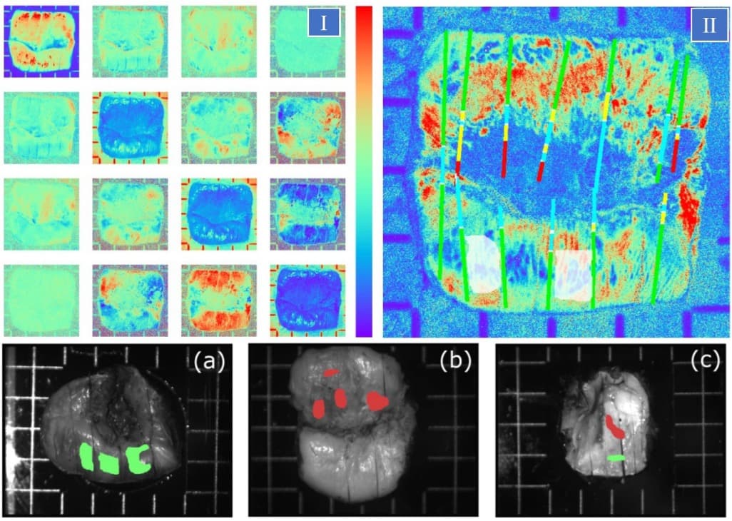
Cervical intraepithelial neoplasia (CIN) progressively disrupts the normal epithelium of the uterine cervix (loss of cell polarity, nuclear enlargement) and remodels the stromal extracellular collagen matrix. Because polarized light is sensitive to exactly these structural features, Mueller matrix images encode the contrast between precancerous zones and healthy uterine cervix tissue that is not visible in greyscale intensity images.
From the measured Mueller matrix images we derive interpretable diagnostic maps, most notably that of linear retardance, which reflects the organization of stromal collagen. Overlaying histologically labeled cut lines (healthy, low-grade CIN, high-grade CIN, glandular and metaplastic epithelium) onto these maps lets us connect each polarimetric signature directly to a histology ground truth diagnosis.
From Specimen Imaging to Specimen Classification
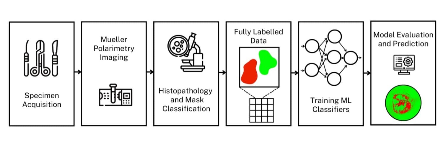
AOPLab has built a polarimetric data post-processing pipeline based on trained neural network classifiers. Excised cervical specimens are measured with the multi-wavelength wide-field imaging Mueller polarimeter and then sent for histopathology analysis for gold standard diagnosis. Expert annotations are co-registered with the polarimetric images to create a fully labeled dataset of healthy and high-grade CIN regions, pixel by pixel.
This labeled dataset makes objective, quantitative classification possible: instead of relying on an expert's visual judgment of live colposcopy images, machine learning models are trained on ground-truth-verified polarimetric features and can then be used for diagnostic evaluation of polarimetric images from unseen uterine cervices.
Machine Learning-Based Detection of Cervical Intraepithelial Neoplasia: Diagnostic Performance
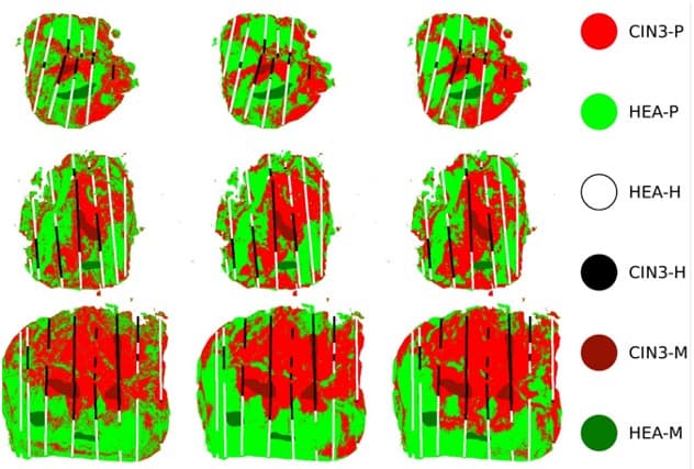
Using this annotated dataset, we compared several machine learning approaches: a decision tree, a multi-layer perceptron, and a 1D convolutional neural network, for pixel-wise classification of healthy versus high-grade CIN3 zones of cervical tissue. Under a stratified train–test split, the best models reached AUC and accuracy around 0.96–0.99. The more demanding leave-one-patient-out validation, which tests true generalization to new patient data, remains the key challenge and the focus of ongoing work.
The predicted maps can be compared side by side with the histological ground truth masks, and the agreement along the diagnostic cut lines gives an interpretable, whole-sample accuracy metric. This ex vivo study, carried out within the BiCPIC and POLANNs projects in collaboration with the University of Arizona and Victoria University of Wellington, is at the proof-of-concept stage toward an objective, polarimetry-based cervical precancer screening aid.
Cervical Remodelling and Preterm Birth Risk
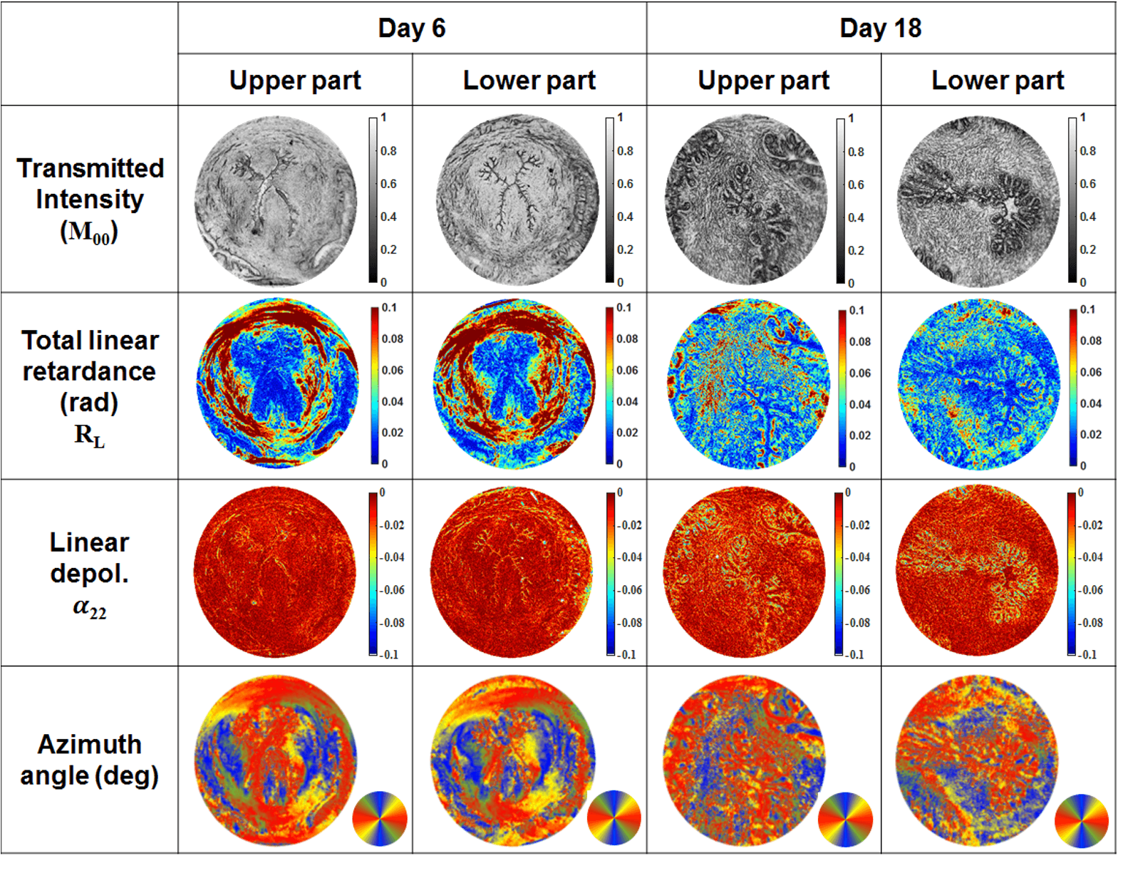
The cervix is a load-bearing structure rich in organized collagen. As pregnancy progresses, it softens and remodels: collagen shifts from an ordered, circumferential arrangement to a disordered one to facilitate delivery of the baby during labor. Premature remodeling is linked to the risk of preterm birth. For this pathology, reliable early markers are still missing. Because polarimetric measurements are sensitive to tissue anisotropy resulting from the presence of arranged collagen fibers, they can directly follow the reorganization of cervical collagen and monitor the risk of preterm birth. In a mouse model of pregnancy, Mueller matrix-derived maps of linear retardance and azimuth change markedly between early and late gestation, pointing toward objective metrics for collagen scoring during pregnancy.
Related Publications
Chae, S., Ajmal, A., Pei, J., Sanchez, A., Boonya-ananta, T., Rodriguez, A., Novikova, T., Ramella-Roman, J. C.
Gestational Stage Prediction from Cervical Tissue Analysis Using Imaging Mueller Polarimetry Data
·2026
Chae, S., Giammattei, D., Ajmal, A., Pei, J., Sanchez, A., Boonya-ananta, T., Rodriguez, A., Ramella-Roman, J. C., Novikova, T.
Journal of Microscopy·2026
Robinson, D., Kleijn, W. B., Novikova, T., Doronin, A.
Diffusion modelling of polarimetric cervical tissue properties
Proc. SPIE 13854, 1385402·2026
Chae, S., Ajmal, A., Rodriguez, A., Boonya-ananta, T., Pei, J., D'Aquino, M., Sanchez, A., Ramella-Roman, J. C., Novikova, T.
Proc. SPIE 14098, Tissue Optics and Photonics IV (SPIE Photonics Europe)·2026
Chae, S., Rodriguez, A., Boonya-ananta, T., Ajmal, A., Pei, J., Sanchez, A., Ramella-Roman, J. C., Novikova, T.
Gestational stage prediction from cervical tissue analysis using imaging Mueller polarimetry data
Proc. SPIE 14098, Tissue Optics and Photonics IV, 140981D (SPIE Photonics Europe)·2026
Robinson, D., Kleijn, W. B., Novikova, T., Doronin, A.
Uncertainty-aware Mueller matrix polarimetry using Gaussian process pipeline
Proc. SPIE 13322, Polarized Light and Optical Angular Momentum for Biomedical Diagnostics 2025, 133220B·2025
Robinson, D., Kleijn, W. B., Hoong, K., Doronin, A., Rehbinder, J., Vizet, J., Pierangelo, A., Novikova, T.
Polarimetric imaging for cervical pre-cancer screening aided by machine learning: ex vivo studies
Journal of Biomedical Optics, 28(10), 102904·2023
Pierangelo, A., Novikova, T., Rehbinder, J., Nazac, A., Vizet, J.
Mueller Polarimetric Imaging for Cervical Intraepithelial Neoplasia Detection
Polarized Light in Biomedical Imaging and Sensing, Springer·2023
Gonzalez, M., Roa, C., Jimenez, A., Gomez-Guevara, R., Du Le, V. N., Novikova, T., Ramella-Roman, J. C.
Proc. SPIE 11963, Polarized Light and Optical Angular Momentum for Biomedical Diagnostics II, 119630B·2022
Lee, H. R., Saytashev, I., Du Le, V. N., Mahendroo, M., Ramella-Roman, J. C., Novikova, T.
Mueller matrix imaging for collagen scoring in mice model of pregnancy
Scientific Reports, 11(1), 15621·2021
Kupinski, M., Boffety, M., Goudail, F., Ossikovski, R., Pierangelo, A., Rehbinder, J., Vizet, J., Novikova, T.
Polarimetric Measurement Utility for Pre-cancer Detection from Uterine Cervix Specimens
Biomedical Optics Express, 9(11), 5691–5702·2018
Novikova, T.
Optical techniques for cervical neoplasia detection
Beilstein Journal of Nanotechnology, 8, 1844–1862·2017
Rehbinder, J., Haddad, H., Deby, S., Teig, B., Nazac, A., Novikova, T., Pierangelo, A., Moreau, F.
Ex vivo Mueller polarimetric imaging of the uterine cervix: a first statistical evaluation
Journal of Biomedical Optics, 21(7), 071113·2016
Pierangelo, A., Nazac, A., Benali, A., Validire, P., Cohen, H., Novikova, T., Haj Ibrahim, B., Manhas, S., Fallet, C., Antonelli, M.-R., De Martino, A.
Polarimetric imaging of uterine cervix: a case study
Optics Express, 21(12), 14120·2013